Specialized Diagnostic Testing
Lake Oconee Eye Care offers the latest in diagnostic technology. This instrument takes 20 diagnostic measurements analyzing thousands of data points to give the most detailed analysis of the visual system available.
Ocular Coherence Tomography (OCT)
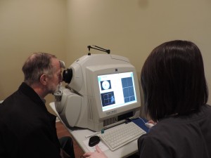 OCT is used to confirm the diagnosis or monitor the progression of a wide variety of eye diseases and conditions. Some of those include, glaucoma, retinal detachment, diabetic and hypertensive retinopathy and wet macular degeneration to name a few. At Lake Oconee Eye Care Vision Source, our Eye care providers believe it is essential to have an OCT on the premises in order to detect those retinal problems while the patient is in the office, so changes in their patient’s eyes can be detected immediately and appropriate referrals can be made when necessary.
Fundus Photography
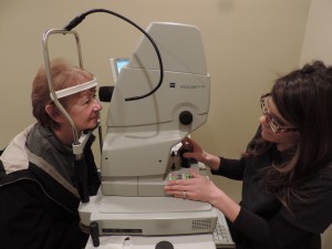 Often it is necessary to take photos of the back of your eyes in order to document original findings of disease or to follow changes that may occur over time, such as trends or changes in the optic nerve in glaucoma or hemorrhages of a branch retinal vein occlusion. Patients may have suspicious pigment in the retina that we document over time to check for changes in size and other properties to insure this is not ocular melanoma. Whatever the reason for which it is needed, our state of the art fundus camera, will document any conditions of the retina that our patients may have.
Epic Refraction System OPD & Corneal Topography
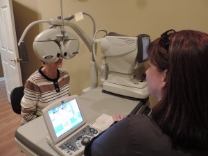 The EPIC refraction system with OPD is a state of the art refraction system that measures both the daytime and nighttime prescription through wavefront technology. It is the first and only system of its kind. It is non-invasive, fast and is less frustrating for the patient than traditional refraction. At the same time your prescription information is being gathered, the instrument is also taking a picture of the anterior segment of your eye as well as mapping the corneal surface (corneal topography) to check for surface irregularities and aberrations that may be preventing clear vision that had previously been undetected and may be the reason for poor vision (instead of what you always thought was a problem with your glasses or contacts!). The multifunctional EPIC is an amazing tool and the most advanced technology available, and our doctors consider it an integral part of the comprehensive eye health exam in order for the patient to receive the best care possible.
Macular Risk Genetic Testing
Did you know there is a test that gives the percentage risk of a patient with dry (non-exudative) macular degeneration (AMD) of converting to wet (exudative) AMD over a two, five and ten year period of time? The test is as simple as a cheek swab that is done while sitting in the exam chair at the conclusion of your eye examination. The lab processes the sample and the results are reviewed with our patients in approximately six weeks. The results of the test, along with the stage of a patient’s current macular degeneration, dictate the appropriate treatment protocol including how often to be dilated and have the maculae photo-documented, special nutritional supplements tailored to your genetic code (provided by the lab results) and appropriate referrals when necessary.